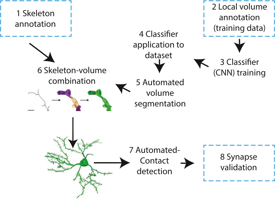
SegEM offers you a workflow for the image analysis of 3D electron microscopy images for connectomics. All SegEM code is availible via a repository on github. The code base on github is continuously improved, we therefore recommend using the code version on github. Alternatively, you can use the code as contained in the supplementary material of the SegEM publication at Neuron.
To benchmark progress in image processing for connectomics, SegEM also provides the SegEM 3-D EM segmentation challenge: large volumes of training data, skeleton-based training data, code to compute the required metrics, and a regularly updated ranking table. To participate, download the SegEM challenge package, and submit your results to SegEMchallenge@brain.mpg.de.

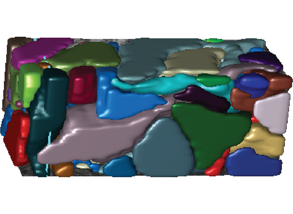
Some visualizations of the capabilities of SegEM. Feel free to use for educational and scientific purposes. © Berning, Boergens, Helmstaedter, Max Planck Institute for Brain Research.
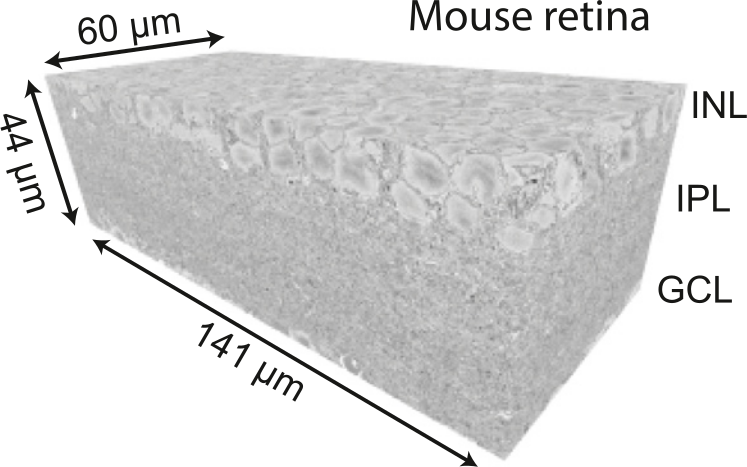
Get access to the large-volume 3D raw data, classifier output, segmentations and skeleton reconstructions used in the SegEM publication. Also, the successful CNNs can be downloaded and directly applied to novel EM datasets:
SegEM was developed at the Max Planck Institutes of Neurobiology (2011-2014) and Brain Research (2014 onwards). It is maintained by the Connectomics department at the Max Planck Institute for Brain Research, Frankfurt. All research was funded by the Max Planck Society.